Picture Of Forearm Tendons / Forearm Pain: A New Treatment For An Old Problem - DRUM ... : Forearm flexor muscles, labeled drawing.
Picture Of Forearm Tendons / Forearm Pain: A New Treatment For An Old Problem - DRUM ... : Forearm flexor muscles, labeled drawing.. Pain, swelling, and redness of the forearm are the most commonsymptoms of the condition. Middle of the lateral surface of the body the radius n. Forearm tendinitis is a painful condition caused by inflammation of a tendon, i.e., a sinew that connects muscle to bone. Four superficial, one intermediate and three deep muscles occupy the anterior forearm. Both tendons and ligaments are dense regular connective tissue, because of its two properties:
The two most common types of tendinitis are on the inside or outside of your elbow. Both tendons and ligaments are dense regular connective tissue, because of its two properties: Forearm flexor muscles, labeled drawing. We can tell this is a ventral view of the forearm because we can see the palmar aponeurosis (a thin, tendinous sheath that is only on the palmar side of the hand) and. The picture above is an example of a great stretch for the inner forearm muscles and tendons, do this stretch before during and after you climb both indoor and outdoor.
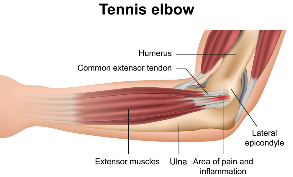
This picture also contains other parts such extensor carpi radialis long, medial epicondyle of humerus, lateral epicondyle of humerus, olecranon of the ulna, extensor carpi ulnarıs, extensor dıgıtorum, flexor carpi ulnaris, extensor retinaculum, tendons of extensor digitorum and so on.
Forearm tendonitis is inflammation of the tendons of the forearm. Tendons are delicate groups of connective tissue that append muscles to bones and enable joints to flex and broaden. Many people will relate to bicep tendinitis of the upper bicep location where the tendon and muscle for the inner bicep, i did the exact same thing, just mirroring the tape on the outside (see picture). Both tendons and ligaments are dense regular connective tissue, because of its two properties: This picture also contains other parts such extensor carpi radialis long, medial epicondyle of humerus, lateral epicondyle of humerus, olecranon of the ulna, extensor carpi ulnarıs, extensor dıgıtorum, flexor carpi ulnaris, extensor retinaculum, tendons of extensor digitorum and so on. 12 photos of the forearm tendon anatomy picture. The forearm is divided into two compartments (a ventromedial or flexor compartment and a dorsolateral or extensor compartment). The superficial group (pronator teres, flexor carpi radialis, plamaris longus and. It hurts, not just when you lift or exercise, but also when you do everyday tasks, even something as basic as typing or moving the mouse on your computer. Pronation of forearm, flexes elbow. Arrangement of forearm muscles and tendons in the wrist. The achilles tendon is the largest and strongest tendon in the human body. Medial supracondylar ridge of humerus i.
The muscle of the common extensor tendon that is nearest this side of the arm is the extensor carpi ulnaris, which attaches to the proximal end of the fifth metacarpal, or the palm bone beneath the pinky. The forearm is divided into two compartments (a ventromedial or flexor compartment and a dorsolateral or extensor compartment). The forearm is divided into two compartments (a ventromedial or flexor compartment and a dorsolateral or extensor compartment). You may be able to treat forearm tendonitis with rest and rice therapy. Tendons are similar to ligaments;
Forearm tendonitis is a condition in which the tendons in the forearm become inflamed and painful.
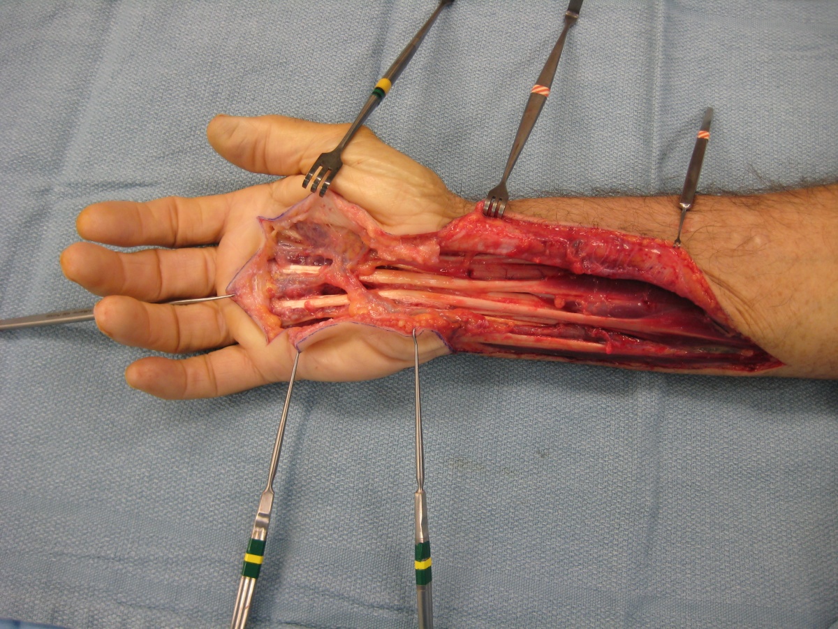
Arrangement of forearm muscles and tendons in the wrist. The posterior view of the muscles of the human forearm. (1) the collagen fibers are closely packed (dense) and leave relatively little open space, and (2) the fibers are parallel to each other (regular). They are shown in the illustration below. Read about symptoms, testing, treatment, and recovery from a ruptured achilles tendon. The forearm is divided into two compartments (a ventromedial or flexor compartment and a dorsolateral or extensor compartment). Read or download of tendons in forearm for free in forearm at. Forearm tendinitis is a painful condition caused by inflammation of a tendon, i.e., a sinew that connects muscle to bone. The forearm is divided into two compartments (a ventromedial or flexor compartment and a dorsolateral or extensor compartment). It hurts, not just when you lift or exercise, but also when you do everyday tasks, even something as basic as typing or moving the mouse on your computer. Forearm pain from muscle or tendon injuries can be quite debilitating. 12 photos of the forearm tendon anatomy picture. Find the perfect extensor tendon stock photos and editorial news pictures from getty images.
Picture 1 shows the achilles tendon and its attachment to the heel bone. Forearm pain from muscle or tendon injuries can be quite debilitating. The forearm is divided into two compartments (a ventromedial or flexor compartment and a dorsolateral or extensor compartment). Both are made of collagen. Read about ruptured tendon symptoms, treatment, and prognosis, whether it's an achilles tendon rupture or the tendon rupture is in the quadriceps, finger, ankle, hand, wrist, elbow, shoulder, knee, or anywhere else in the.
Medial supracondylar ridge of humerus i.
Forearm muscles (common flexor tendon). Injuries the common conditions within the tendons throughout the elbow joint comprising of the tennis elbow, and the golfer's elbow, which occur from an overuse injury to the tendons or result from. Forearm tendonitis is aggravation of the tendons of the lower arm. The muscle of the common extensor tendon that is nearest this side of the arm is the extensor carpi ulnaris, which attaches to the proximal end of the fifth metacarpal, or the palm bone beneath the pinky. Your walls are a reflection of your personality, so let them speak with your favorite quotes, art, or designs printed on our the forearm muscles and tendons become damaged from overuse — repeating the same motions again and again. The parallel arrangement of fibers is an adaptation to the fact that. Picture 1 shows the achilles tendon and its attachment to the heel bone. Read about ruptured tendon symptoms, treatment, and prognosis, whether it's an achilles tendon rupture or the tendon rupture is in the quadriceps, finger, ankle, hand, wrist, elbow, shoulder, knee, or anywhere else in the. We can tell this is a ventral view of the forearm because we can see the palmar aponeurosis (a thin, tendinous sheath that is only on the palmar side of the hand) and. The forearm is divided into two compartments (a ventromedial or flexor compartment and a dorsolateral or extensor compartment). Tendons are similar to ligaments; Tendons are delicate groups of connective tissue that append muscles to bones and enable joints to flex and broaden. Picture of the achilles tendon.


Komentar
Posting Komentar